What is acute myeloid leukemia (AML)?

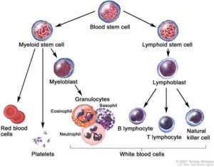
AML is a cancer of the blood in which the bone marrow makes too many immature myelocytes (white blood cells). All blood is made in the bone marrow where blood cells come from stem cells. Acute myeloid leukemia (AML) is a cancer of the white blood cells, where too many immature abnormal white blood cells, called “blasts”, are found in the bone marrow. There are numerous kinds of AML which were originally classified based on their appearance under the microscope. These blasts crowd out the normal blood cells in the bone marrow and spread to the blood. They can also spread to the brain, spinal cord, and/or other organs. Myeloblasts do not work like normal myeloid cells and are not able to fight infection. As the number of leukemia cells increases in the blood and bone marrow, there is less room for healthy red blood cells, and platelets which leads to anemia, and easy bleeding.

What are signs of childhood AML?

  • Fever.
  • Masses in the skull or other parts (chloroma)
  • Easy bruising or bleeding.
  • Bone or joint pain.
  • Painless lumps in the neck, underarm, stomach, or groin.
  • Pain or feeling of fullness below the ribs.
  • Weakness, feeling tired, or looking pale.

What causes AML?

Nobody knows for sure, but commonly there is nothing in the environment or diet that caused this. Some cases of childhood AML are associated with genetic disorders such as Down Syndrome, Fanconi Anemia, and Kostmann Syndrome. Most cases and are not inherited so other children in the family should not be at risk, your doctor should be able to confirm that. It is not infectious so it cannot be passed onto other children or playmates.

How is AML diagnosed?

We may suspect the diagnosis from the blood count but the tests described below will confirm it and find out if leukemia has spread to other parts of the body e.g. spinal fluid

  • Physical exam and history: An exam of the body to check or signs of disease, such as lumps or anything else that seems unusual e.g. testes enlargement.
  • Blood tests: such as complete blood count (CBC) which tells the number and type of each blood cell, and blood chemistry studies because certain substances may be increased in acute leukemia.
  • Bone marrow aspiration and biopsy: Removal of liquid bone marrow and a tiny piece of bone by inserting a needle into the hipbone while the child is sedated. A pathologist views the marrow sample under a microscope to look at the leukemia cells. Special tests done include flow cytometry to look at markers on the surface of the cells to confirm the diagnosis and subtype of AML and genetic analysis of the leukemia cells.
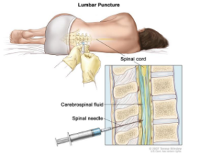
  • Lumbar puncture: The removal of a teaspoon of cerebrospinal fluid (CSF) by placing a needle between two bones in the lower spine while the child is sedated. CSF is reviewed under a microscope for leukemia, and chemotherapy is injected at the same time. This is repeated throughout the entire course of treatment so that leukemia cells don’t hide there.

Is childhood AML curable?

Childhood AML is more curable than in adults, and long-term cure rates with modern day chemotherapy may be as high as 80% if your child is in a low-risk group and receives up-todate treatment. Cure rates vary widely depending on which risk group he or she is in. For example, M3/APL responds so well to specific targeted drugs that it is discussed in a separately.

Chances of long-term cure depend on knowing with certainty the subtype and genetics of the AML your child has. The French-American-British (FAB) Classification System listed below was based on how leukemic cells look under the microscope. This has now been replaced by the World Health Organization (WHO) system which includes the genetic changes in the leukemia cells.

  • M0/M1: Acute myeloblastic leukemia with or without minimal differentiation
  • M2: Acute myeloblastic leukemia with differentiation
  • M3: Acute promyelocytic leukemia (APL)
  • M4/M4Eo: Acute myelomonocytic leukemia (AMML) without or with eosinophilia
  • M5a/M5b: Acute monocytic leukemia (AMoL) with or without differentiation
  • M6: Acute erythroid leukemia (AEL).
  • M7: Acute megakaryocytic leukemia (AMKL)


Low risk: Core-binding factor (CBF) AML accounts for 20% of childhood AML and includes cases with t(8;21) RUNX1-RUNX1T1 and inv(16) CBFB-MYH11 fusion genes, with a 5-year survival of around 80% with standard chemotherapy.

High risk: Abnormalities involving chromosomes 5 and 7 (especially monosomy) account for <5% childhood AML with a 5-year survival of <30% even with bone marrow transplant (BMT). FLT3-ITD mutation is found in 5-10% childhood AML and may indicate a worse prognosis.

Intermediate/uncertain risk: KMT2A (MLL) gene rearrangements account for 20% of childhood AML, with the majority t(9;11) MLLT3-KMT2A and a 5-year survival of around 50% but with >50 other fusion partners, an exact prognosis depends on the precise rearrangement. In addition, a special bone marrow test done at the end of induction treatment (MRD test) with a goal of <0.1% may help guide treatment for patients in the intermediate risk group.

Since there may be multiple genetic changes in the AML cells, expert review by an experienced pathologist and clinical team is needed. Your doctor will use this information to determine your risk group and plan the chemotherapy treatment with the best chance of cure.

What treatment will my child receive?

In the initial stage of treatment called Induction, we try to remove all visible signs of leukemia and allow normal blood cells to be restored, which is called remission and is usually achieved by the end of induction. Cancer fighting medicine (chemotherapy) is given as a standard combination. Chemotherapy is injected into a vein or muscle to reach cancer cells throughout the body. Chemotherapy is also placed directly into the cerebrospinal fluid (intrathecal). Your doctor will use information gathered during the first month of treatment to determine your final risk group, and what further treatment your child should receive.

In AML chemotherapy drugs are given together at high doses in cycles, each of which lasts 3-4 weeks, to increase the chances of killing all the leukemia cells and getting a cure. The majority of patients will require several weeks inpatient in the hospital in order to receive the chemotherapy or due to its side effects of low blood counts and infections. Modern-day AML chemotherapy protocols treat with 4-5 cycles of intensive chemotherapy over 5 months.

All people who receive cancer treatment are at risk of having side effects, some of which are mild but others of which can be life-threatening In addition to killing tumor cells, chemotherapy can damage normal tissue. Side effects usually get better when the medication is stopped but sometimes last a long time or never go away. Common side effects include nausea, vomiting, hair loss, low blood counts and fatigue (tiredness). Drugs are given to prevent nausea and vomiting. Hair loss is usually temporary but very rarely it may be permanent. Low blood counts may require transfusion or increase the risk of infection. Chemotherapy side effects are listed on the sheets your doctor will give you, these vary depending on the medication. It is important to remember that the treatment given is essential for cure and most patients will have very few of the side effects listed.

Radiation treatment is not needed unless there is evidence of AML masses outside the bone marrow (chloroma). The presence of such masses does not reduce the curability of the AML.

Will my child need bone marrow transplantation (BMT)?

Low and intermediate risk patients should not need BMT, but many patients with high risk will benefit. Uncertainty has been created in the past by differing definitions of risk categories used by different cooperative groups, nevertheless consensus is emerging that for high-risk patients with a matched family donor, BMT helps improve the likelihood of cure

What dietary and what other precautions should I take?

Chemotherapy suppresses your child’s immune system so infections are a major concern. We place diet and activity restrictions throughout treatment. We prohibit any street/outside food and recommend home food be thoroughly cooked. No special foods will help with cure but we ban vitamin or nutritional supplements that might interfere with chemotherapy. Visits to movie theaters, malls, weddings, or other crowded places and plane, bus or railway travel are all banned. You should contact your doctor immediately if in spite of precautions your child develops a fever >100.4F, diarrhea, cough, vomiting, abdominal pain, headaches or looks unwell.

Sample treatment protocol for child with AML

Methods for Giving Chemotherapy

  • IV – Drug is given using a needle or tubing inserted into a vein. Drugs can be given rapidly over a few minutes (“push”) or slowly over minutes or hours (“infusion”).
    over a few minutes (“push”) or slowly over minutes or hours (“infusion”).
  • IM – Drug is given into a muscle using a needle.
  • IT – Drug used to treat the brain and spinal cord is given using a needle inserted through the back into the fluid surrounding the spinal cord.
  • SC – Drug is given by subcutaneous injection

Standard Treatment Table (Low-risk patients)

Induction I and II are meant to kill as many of the leukemia cells as possible so the disease goes into remission, each cycle lasts about 4 weeks.

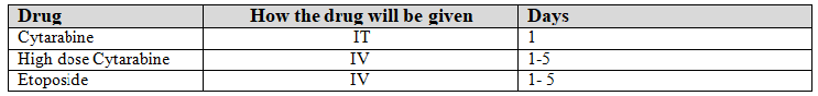
*Subjects with leukemia in the CNS (CNS3) also get IT Ara-C twice weekly until CSF is clears.

Intensification I is to kill all remaining leukemia cells and lasts about 4 weeks.

Intensification II is to kill all remaining leukemia cells and lasts about 4 weeks.