What is acute lymphoblastic leukemia (ALL)?
ALL is a cancer of the blood in which the bone marrow makes too many immature lymphocytes (white blood cells). All blood is made in the bone marrow where blood cells come from stem cells. Acute lymphoblastic leukemia (ALL) is a cancer of the white blood cells, where too many immature abnormal white blood cells, called “blasts”, are found in the bone marrow. Depending on which kind of lymphoblast is in excess it may be pre-B cell leukemia, or (more rarely) T-cell leukemia. These blasts crowd out the normal blood cells in the bone marrow and spread to the blood. They can also spread to the brain, spinal cord, and/or other organs of the body. Lymphoblasts do not work like normal lymphocytes and are not able to fight infection. As the number of leukemia cells increases in the blood and bone marrow, there is less room for healthy red blood cells, and platelets which may also lead to anemia, and easy bleeding.

What are signs of childhood ALL?
- Fever.
- Large mass in chest (T-cell only)
- Easy bruising/bleeding
- Bone or joint pain.
- Weakness, feeling tired, or looking pale
What causes ALL?
Nobody knows for sure, but commonly there is nothing in the environment or diet that causedthis. It is typically not inherited so other children in the family should not be at risk and it is not infectious so it cannot be passed onto other children or playmates.
How is ALL diagnosed?
We may suspect the diagnosis from the blood count but the tests described below will confirm it and find out if leukemia has spread to other parts of the body e.g. spinal fluid
- Physical exam and history: An exam of the body to check or signs of disease, such as lumps or anything else that seems unusual e.g. testes enlargement.
- Blood tests: such as complete blood count (CBC) which tells the number and type of each blood cell, and blood chemistry studies because certain substances may be increased in acute leukemia.
- Chest x-ray: to see if leukemia cells have formed a mass in the middle of the chest.
- Bone marrow aspiration and biopsy: Removal of liquid bone marrow and a tiny piece of bone by inserting a needle into the hipbone while the child is sedated. A pathologist views the marrow sample under a microscope to look at the leukemia cells. Special tests done include flow cytometry to look at markers on the surface of the cells to confirm the diagnosis and subtype of ALL and genetic analysis of the leukemia cells.

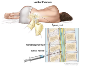
- Lumbar puncture: The removal of a teaspoon of cerebrospinal fluid (CSF) by placing a needle between two bones in the lower spine while the child is sedated. CSF is reviewed under a microscope for leukemia, and chemotherapy is injected at the same time. This is repeated throughout the entire course of treatment so that leukemia cells don’t hide there.
Is childhood ALL curable?
The treatment for T-cell and B-cell ALL is similar, and long-term cure rates with modern day chemotherapy are 80% or higher provided your child is in the correct risk group and receives up-to-date treatment. Cure rates vary depending on the risk factors described below and your doctor will be able to give you a better estimate after taking those all into account.
What are risk Groups in ALL?
Risk groups tell us how likely the ALL is to come back without adequate treatment and are usedto plan chemotherapy. There are three risk groups in childhood ALL and the final risk group isdetermined by your doctor:
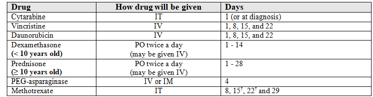

 *Since methotrexate decreases folic acid, replacement with leucovorin will be given starting 42 hours after each dose.
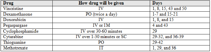
 *For the first 4 cycles (one year), patients get an extra dose on Day 29.